Article for the “Bio/Mol/Text” competition: “Demon by the bed”, “incubus sitting on the chest”, “sleepy stupor” - all these are not just terms of mythology, but a complex phenomenon, which is based on many physiological processes. It has long been colorfully described from a philistine point of view, but little studied by science - this is exactly the impression sleep paralysis (SP) leaves about itself. In our article, we will delve into the physiological basis of the phenomenon in which almost all skeletal muscles lose the ability to move, but the mind remains clear, and we will try to prove that science has not stood still for a long time in studying the mechanisms of sleep paralysis.
Competition “Bio/Mol/Text”-2020/2021
This work was published in the “Free Topic” category of the “Bio/Mol/Text” competition 2020/2021.
The general partner of the competition is the annual biotechnology conference BiotechClub, organized by the international innovative biotechnology company BIOCAD.
The sponsor of the competition is SkyGen: a leading distributor of life science products on the Russian market.
Competition sponsor: the largest supplier of equipment, reagents and consumables for biological research and production.
"Book" sponsor of the competition - "Alpina Non-Fiction"
Imagine opening your eyes just before dawn, trying to roll over in bed, and suddenly realizing that you are completely paralyzed. Lying helplessly on your back and unable to scream for help, you notice sinister figures lurking in your bedroom. As they approach the bed, your heart begins to beat faster and you feel like you're suffocating. You hear a buzzing sound and feel electrical impulses throughout your body. After a moment, the visions disappear and you can move again. Horrified, you think about what just happened. The above experience is an episode of sleep paralysis accompanied by hypnopompic hallucinations [1]. This frightening description has been interpreted in various cultural contexts since the time of the ancient Greeks, with a variety of spiritual and supernatural explanations ranging from witchcraft and evil spirits to aliens [2], [3]. Modern medical explanations of SP are not so colorful, but they help us understand the problem much more deeply.
The artist Henry Fuseli dedicated a series of four paintings to this phenomenon - “Nightmare”. The embodiment of the nightmare is personified by the dissolute demon of the mythology of medieval legends - an incubus sitting on the chest of a young girl. The tone of the picture evokes a depressing feeling, plunging the observer into an atmosphere of hopelessness and horror, making one feel the full severity of the charming girl’s condition under the pressure of a “sleepy stupor.”
Can old witch syndrome be cured?
Since the phenomenon cannot be attributed to diseases or even sleep disorders, there are no methods for treating sleep paralysis. Every ninth inhabitant of the globe experiences such an experience at least once in his life; there is no need to consult a doctor, unless this condition begins to haunt you with enviable regularity, causing mental disorders or other pathologies. You need to see a doctor if:
- you have frequent nightmares;
- sleep paralysis is accompanied by vivid hallucinations;
- you are depressed;
- you suffer from severe headaches;
- you suffer from frequent tachycardia;
- the condition is accompanied by or causes panic attacks.
For all others, treatment for sleep paralysis is not required. It is recommended to treat the condition as adequately as possible and not try to get out of it too quickly. If you encounter this phenomenon, do not make sudden movements and maintain a sober mind.
In order to calm down, you need to control your breathing, try to take as deep a breath as possible, and as you exhale, say a word, a sound, or simply shout. Don’t be afraid that this will wake up your household, because your throat muscles will also be relaxed, which means the sound will come out very quiet. But your consciousness will be able to get involved in the work and disperse the manifestations of paralysis.
Frequent blinking is a great way to get rid of sleep paralysis and its consequences. Open and close your eyelids, thereby bringing yourself out of your stupor. Mental arithmetic helps a lot. And simply switching to the perception of reality can also be useful, since it distracts from the feeling of physical helplessness, which gradually fades. For believers, prayer can be an excellent “medicine”. The effect of self-hypnosis after reading lines addressed to God will help drive away nightmares and give a feeling of protection.
It is extremely important to avoid stress in everyday life. Very often, those who constantly experience psychological stress experience sleep paralysis.
We are talking about people who work in stressful positions or are forced to constantly risk their health, about students, workaholics and perfectionists suffering from “excellent student syndrome.”
Each case has its own answer to what causes sleep paralysis. A guaranteed method to cope with this unpleasant phenomenon is a correct diagnosis and treatment of the root cause. More often we are talking about eliminating mental discomfort provoked by the patient’s environment.
Conversations with a psychologist, relaxing massage, hypnotherapy, water procedures and other means make it possible to almost completely relieve the syndrome. It is worth understanding that if your parents or grandparents also often had sleep paralysis, you will also suffer from them with a high degree of probability
In this case, it is especially important to get enough sleep, lead a healthy lifestyle and get rid of stress.
Follow the rules of sleep hygiene: regularly ventilate the bedroom, sleep on a comfortable mattress, use pleasant bedding, and give your body moderate exercise.
Sometimes sleep paralysis can be triggered by loud noises, such as an alarm clock. Therefore, to avoid troubles, set yourself a soft melody to wake up, or even better, learn to wake up without an alarm clock
Section 1. Epidemiology of sleep paralysis
According to studies conducted by American and Belarusian scientists, the prevalence of sleep paralysis varies among the mentally healthy population, mainly from 5 to 30%, but according to some data it can reach 62% [1]. Thus, the majority of respondents note single or rare episodes, but for some, paralysis recurs [4]. The average age of onset of the first episode of the phenomenon is 17±1 years [5]. The phenomenon is more often observed in females (18.8%) than in males (15.7%) [2]. Rates may also vary by ethnicity. Here, the prevalence of the phenomenon is highest among people of the Negroid race, and representatives of the Asian race are least susceptible to sleep paralysis [6]. Among people belonging to the Caucasian race, the phenomenon gained the greatest “popularity” among students and middle-aged people. However, it currently seems difficult to identify a reliable dependence on ethnicity among the entire population, since researchers cannot find a sufficient number of subjects of pure race [2].
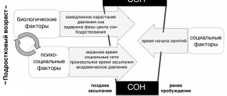
This condition can be inherited due to a mutation in the PER2 gene, which is responsible for the manifestation of circadian rhythms in humans [7]. This gene belongs to the Per gene family and is one of the key biological clock genes. A clear circadian rhythm of its expression in neurons of the suprachiasmatic nuclei has been demonstrated. Genes in this family encode components of the circadian rhythms of locomotor activity, metabolism, and behavior. In humans, a mutation in the PER2 gene has been shown to cause familial advanced sleep phase syndrome (FASPS) [8].
A few centuries ago, at the end of the Middle Ages, scientists began to think about the mechanisms of sleep paralysis. By this time, enough descriptions of this phenomenon had already accumulated. Each nation explained this condition in its own way: some people believed in the existence of witches, others in shamans who took lives, while others believed that demons “have fun” this way. The first documented case of paralysis was described by the Dutch physician Isbrand van Diemerbroek in a 17th century treatise [9]. The author recounted the story of a fifty-year-old physically and mentally healthy woman experiencing bouts of sleep paralysis: “When she tried to sleep, she sometimes believed that the devil was lying and holding her. Sometimes she would be suffocated by a large dog or a thief lying on her chest.” The author wrote that the subject could not even move to drive away the imaginary guests. Since that time, the external manifestations of paralysis have not changed, but scientists were able to describe the phenomenon from a scientific point of view.
What do the statistics show?
The question of what causes sleep paralysis is under study. According to general statistics, approximately 7% of the population has experienced sleep paralysis at least once during their life. Students are more susceptible to it (28.3%). It has been hypothesized that the nature of such muscle paralysis may be due to irregular sleep or prolonged exposure to stress.
Data obtained from a study of the health of students show that 75% of students who experienced a state of sleep paralysis had at least one case of hallucinations at this moment. About 10% of respondents reported a frequency of hallucinatory visions of 3 or more times. 90% of students who were in a state of sleep atony experienced a strong feeling of fear and horror when falling asleep.
This sleep disorder occurs in the fairer sex 1-2% more often than in the stronger sex. Since the difference is insignificant, it means that the risk of developing sleepy stupor does not depend on gender. Sleep paralysis can also occur in children.
Patients with psychiatric diagnoses usually suffer from insomnia atony: it developed in 31.9% of such patients. Among patients diagnosed with panic disorder syndrome, sleepy stupor occurred in 35%.
Section 2. Neurophysiological basis for the occurrence of the phenomenon
2.1. Mechanisms of transition to REM sleep
The structure of our sleep includes two alternating phases: the fast REM phase (rapid eye movement phase) and the slow NREM phase (non-rapid eye movement sleep, i.e. sleep without rapid eye movements). slow sleep). In this article, we will discuss the first of these phases in more detail, since it plays a key role in the occurrence of the phenomenon of sleep paralysis. This phase is also called the paradoxical phase, as it is characterized by increased brain activity, increased cerebral blood flow and increased serotonin levels, which is accompanied by increased blood pressure and heart rate; rapid and shallow breathing is noted, as well as intense movements of the eyeballs and contractions of facial muscles. Dreams usually occur during REM sleep.
One theory suggests that REM sleep is initiated by small clusters of cells located in the pons and medulla oblongata. The main mediators of these structures are acetylcholine and glutamate [10]. Groups of these cells are not arranged into a single “center” of REM sleep, but work as individual components of a wide network (Fig. 1) [11]. For example, activation of locus coeruleus (LCα) neurons leads to the development of muscle atonia; rapid eye movements are initiated by neurons of the reticular formation (RF), located near the abducens nucleus (PAb); muscle contractions occur when neurons in the nucleus giant cell are activated [11], [12].
Activation of a certain group of brain stem cells ( red ovals ) causes a change in the corresponding indicators of the body during the REM sleep phase, which are recorded in the form of curves (Fig. 1, right column). For example, EEG signs of cortical activation during REM sleep are realized due to the simultaneous activation of midbrain RF (MRF) neurons and magnocellular nucleus (MN) neurons in the medulla oblongata; generation of the theta rhythm in the hippocampus is initiated by neurons of the pontine oral reticular nucleus (PO); an increase in brain temperature and cyclical fluctuations in the parameters of the cardiorespiratory system are provided by neurons of the parabrachial nucleus (PBN).
Figure 1. Cellular-molecular-network model of the physiological mechanisms of REM sleep formation.
Blue square: LC — neurons of the locus coeruleus, RN — neurons of the raphe nuclei; Green circle: GABA - local GABAergic neurons; Red ovals: MRF - neurons of the midbrain reticular formation, MN - neurons of the magnocellular nucleus of the medulla oblongata, LCα - neurons of the locus coeruleus alpha, PAb - neurons located near the abducens nucleus, PO - neurons of the oral reticular nucleus of the pons, PBN - neurons of the abrachial nucleus; C-PBL—neurons of the posterolateral peribrachial region, Sub C—neurons located under the locus coeruleus; Yellow rectangle: mPRF - medial pontine reticular formation; Red square: PPT - neurons of the pedunculopontine nucleus, LDT - neurons of the posterolateral part of the tegmentum; above the arrow: KR - kainate receptors; Yellow oval: Glutamate - glutamate [13]; Curves on the right (from top to bottom): EEG of a rat in the REM sleep phase, EMG of a rat in the REM sleep phase, EOG of a rat in the REM sleep phase, the theta rhythm of the hippocampus in the REM sleep phase, cyclic oscillations (BP - blood pressure, HR - heart rate, RESP - respiratory rate, BT - body temperature), PGO waves.
[11]
These groups of brainstem cells responsible for triggering REM signals are activated by increased release of cholinergic neurotransmitters, while the amount of aminergic neurotransmitters is either reduced or completely absent. Sources of acetylcholine (Fig. 1, red arrows) are cholinergic neurons of the pedunculopontine nucleus (PPT) and the posterolateral tegmentum (LDT). The sources of aminergic neurotransmitters (Fig. 1, blue arrows) are noradrenergic neurons of the locus coeruleus (LC) and serotonergic neurons of the raphe nucleus (RN). On the membrane of the former there are kainate receptors, which these neurons need to start working. A similar system is formed by both GABAergic and aminergic neurons.
The development of REM sleep begins with increased release of glutamate (Fig. 1, yellow oval); Consequently, the kainate receptors of cholinergic neurons (KR) are activated, which leads to their excitation and increases the release of acetylcholine in each of the REM sleep signal generators (Fig. 1, red arrows). Bidirectional communication of cholinergic neurons of the pedunculopontine nucleus (PPT) and a group of cells of the posterolateral tegmentum (LDT) with neurons of the medial pontine reticular formation (mPRF) is also triggered. Simultaneously with the described processes, local GABAergic cells are also activated (Fig. 1, green circle), which leads to active inhibition of aminergic neurons of the locus coeruleus and raphe nuclei and reduces or completely stops the release of aminergic neurotransmitters in the structures responsible for the generation of REM sleep.
The key role of the aminergic system is to stop its activity in time when cholinergic neurotransmitters are released. To further maintain episodes of REM sleep, increased acetylcholine release in mPRF neurons activates glutamatergic neurons, which continue to release glutamate in the PPT and LDT to maintain the activity of cholinergic neurons. Thus, cholinergic cells PPT and LDT and glutamatergic cells LC and RN form a positive feedback loop to maintain REM sleep cycles [11].
Another mechanism for the occurrence of REM sleep was proposed by Jouvet and Thompson and called it the “activation-synthesis hypothesis.” According to scientists, dreams - just as in the mechanism described above - are the result of nonspecific activation of neurons in the brainstem. But the analysis does not end there, and the signal is further transmitted to the cerebral cortex and subcortical formations for subsequent synthesis of the received information. Scientists have divided all nerve cells into two categories: cholinergic and monoaminergic REM sleep neurons. REM sleep is the result of the reciprocal interaction of these nerve cells [9].
However, the nerve centers for sleep regulation are located not only in the brainstem, but also in the diencephalon. The GABAergic “NREM sleep center” is located in the anterior hypothalamus, and the orexinergic neurons that ensure the process of turning on the “REM sleep center” are located in its middle part (Fig. 2).
Almost simultaneously, two groups of researchers from Japan and the USA discovered two amino acid sequences that were similar to each other, but named them differently [14]. It soon became clear that these were the same substance - the oligopeptides orexin A, containing 33 amino acid residues, and orexin B, consisting of 28 amino acids [15]. The protein proorexin is cleaved from preproorexin, from which orexins A and B are derived. The effect of the latter is determined by two G-protein-coupled metabotropic receptors. OX1R is a type 1 receptor that selectively binds only to orexin A, while OX2R is a type 2 receptor that is nonselective and binds to both orexins. The orexin A receptor is associated only with the Gq subclass of heterotrimeric G proteins, the orexin B receptor is associated with the Gq subclasses or Go and Gi proteins (Fig. 2) [16]. Orexinergic nerve cells interact with locus coeruleus neurons, inhibiting them [14].
Figure 2. Orexins and their receptors.
LC - locus coeruleus, VTA - ventral tegmentum, VMN - ventromedial hypothalamus, DR - raphe nuclei, LDT/PPT - dorsolateral laterodorsal/pedunculopontine tegmentum, BST - bed nucleus of the stria terminalis, PVN - paraventricular hypothalamic nucleus, Arc - semicircular nucleus, TMN - tuberomammillary nucleus, DMH - dorsomedial nucleus, PVT - paraventricular thalamic nucleus.
[16]
2.2. How can disruption of the mechanisms of transition to REM sleep lead to SP?
Research by American scientists at the University of Pennsylvania has shown that the basis of SP is the perseveration of REM activity, that is, a violation of the switch from sleep to wakefulness [2].
- Perseveration (Latin perseveratio - “persistence”, “persistence”) - a stable repetition of something, in this case - REM activity.
During the REM phase of sleep, our brain systems are actively working, as we verified above, but the body remains motionless [6]. But sometimes during this period an awakening occurs, and if the brain structures do not have time to synchronize with each other, paralysis occurs [17]. At the molecular level, awakening is mediated by the following mechanisms. It is believed that the cholinergic and glutamatergic systems are mainly associated with the bioelectrical and behavioral manifestations of the awakening process [18]. The nuclei of the brainstem, responsible for excitation and containing acetylcholine, dopamine, serotonin or norepinephrine, activate the thalamus, hypothalamus, motor neurons of the spinal cord, forebrain and have an inhibitory effect on the ventrolateral preoptic area (GABA, galanin); The hypothalamic and thalamic excitatory centers activate the cortex and arousal-related areas in the basal forebrain and brainstem [19].
One of the most notable aspects of SP is vivid hypnogogic or hypnopompic hallucinations [1]. They can occur during a sudden awakening, along with visions of demonic images, a state of panic, and a feeling of the presence of strangers [20]. A possible explanation for this phenomenon may be that during REM sleep there is a decrease in the activity of the respiratory muscles; this is due to inhibition of motor neurons. During REM sleep, breathing becomes irregular, skeletal muscle hypotension occurs, which leads to a significant decrease in lung ventilation and tidal volume, which leads to excessive accumulation of carbon dioxide in the blood - hypercapnia - and a decrease in the amount of oxygen - hypoxia [21]. In addition, oligodendrocytes, the myelin-producing cells of the central nervous system, are selectively sensitive to hypoxia and sleep fragmentation [22].
- Hypnogic hallucinations are visual illusions that appear before falling asleep.
- Hypnopompic hallucinations are visual illusions of perception that appear after awakening.
A century ago, American neurologist Weir Mitchell described the state of “night paralysis” as follows: “The subject awakens and is aware of his surroundings, but cannot move his muscles; lying there, apparently still sleeping. He is really involved in the struggle for the movement, fraught with acute mental disorder; if he had time to move, the spell would have disappeared instantly” [23].
It is assumed that in addition to the secondary (and sometimes primary) sensory cortex, dysfunction in other areas - the premotor cortex, the cingulate cortex, the subcortical and cerebellar areas - also contributes to the occurrence of hallucinations. The disorder that causes hallucinations is almost always located in areas of the brain associated with sensory systems. It is assumed that compensatory overactivation of brain regions surrounding these pathways is also the cause of hallucinations [24]. However, within seconds to minutes of awakening, the perceptual, cognitive, and motor components of the sleep cycle are synchronized, hallucinations disappear, and mobility is restored when the person is fully awake.
The mechanisms of REM-on and REM-off systems block impulses arriving along incoming sensory fibers, supplying the cortex with internal stimuli that form the content of dreams. They also block the activity of motor neurons in the cortex, thereby immobilizing the dreamer [1].
Thus, when sleep paralysis occurs during a sudden unplanned awakening during REM sleep, eye movements remain unchanged, showing the same high activity, and sensory sensations are clear.
Section 3. Methods for studying sleep paralysis
3.1. Polls
Important methods for studying and diagnosing the phenomenon of sleep paralysis are considered to be a survey and examination of people who have had a similar experience, as well as polysomnography [25], [26].
Surveys are conducted among healthy people who are asked various kinds of questions about their feelings during the period of occurrence of the phenomenon [27]. They mainly evaluate the state of the person’s nervous system during the period of paralysis, the presence of concomitant pathology or increased anxiety, pace of life, place of work or study. The following surveys are often used in research today:
- FISPI - fearful isolated sleep paralysis inventory;
- ISPQ - isolated sleep paralysis questionnaire [28];
- SEQ - sleep experiences questionnaire;
- SPQ - sleep paralysis questionnaire [6];
- USEQ - Unusual sleep experiences questionnaire;
- WUSEQ - Waterloo unusual sleep experiences questionnaire.
A semi-structured interview (FISPI) assesses the frequency of sleep paralysis episodes, the presence of fear and specific hallucinations, dangerous variants of sleep paralysis, and the presence of recurrent episodes. Thanks to this survey, it is possible to conduct a general assessment of distress both during the SP and after over the past 6 months [6].
The ISPQ, created by scientists at the Sacré-Coeur de Montreal Hospital and the University of Montreal, assesses the frequency of episodes of SP using a scale from “never” to “more than 10 times.” Some of the survey questions concern the presence of a sense of someone's presence, which could, for example, be described as follows: “Just before going to bed or after waking up, I felt a presence, not necessarily seeing or hearing anything, in the room where I was lying” (translation from French original) [29].
The USEQ test survey was developed by American scientists to assess the phenomenology of SP. To begin with, respondents are asked an introductory question, after answering which some of the subjects usually stop participating in the survey: “Some people have had experiences when they went to bed or woke up when they could not move their arms or legs, or speak, even if they wanted to. . Have you ever had such an experience? Participants who denied having such feelings stopped completing the questionnaire. The importance of this opening question is that sometimes people can misinterpret the experience. If the survey continues, then the subjects are asked to remember their most vivid episode of paralysis and describe it according to the main characteristics: duration, how long ago the episode was, events accompanying it, etc. The questionnaire also contains questions with answer options on a 4-point scale - from 1 ("never") to 4 ("always") and on a 5-point scale - from 1 ("not at all") to 5 ("a lot") [ 29].
The Waterloo Questionnaire (WUSEQ) also uses the 4-point scale described above to determine the frequency of occurrence of the phenomenon. The brightness and intensity of experiences are assessed in the same way, but in a 7-point range: from 1 (experience of vague signs of a phenomenon) to 7 (experience of a very clear impression of a phenomenon). In total, the questionnaire consists of 42 items [30].
There is also a form of questioning in which the subject shares his experience in the format of a complete description of what was happening to him at the time of sleep paralysis: “Once, during sleep, a sharp unpleasant noise was heard. I seemed to have woken up, but in some other world. Trembling shook from head to toe. Next I felt a strong pressure on my chest; there was a feeling that someone was sitting on top and, trying to strangle me, was pressing me into the bed. It became difficult to breathe. I was overcome with horror and fear. I tried to scream, but in vain; I couldn’t make a single sound. All sorts of thoughts flashed through my head: “I’m going to die,” “I’m already dead,” “I’m in a coma,” etc. What was happening seemed like an eternity, the pressure became unbearable. But suddenly, the trembling suddenly stopped, the noise disappeared” [20].
Despite the fact that many people who have experienced this phenomenon themselves are left with not very pleasant feelings, there are also those for whom the new experience brings positive emotions. Thus, research by a group of Czech scientists showed that more open people are less likely to negatively interpret often anomalous experiences associated with SP, and are more likely to enjoy new sensations and hallucinations [25].
Recommendations suggest that any assessment of SP should be conducted more broadly in the diagnostic context of other psychiatric conditions. For example, using a structured clinical interview - Diagnostic and Statistical Manual of Mental Disorders, 5th edition DSM-5 (Diagnostic and Statistical Manual of mental disorders, fifth edition) [3].
3.2. Polysomnography
The polysomnography method is used to diagnose certain diseases, the manifestation of which can only be noticed during night sleep. This study is painless, practically not felt by the patient and has no contraindications for use [26].
Polysomnography is a complex of studies of changes in the physiological parameters of the body during sleep, which includes: electroencephalography (EEG), electrooculography (EOG), electromyography (EMG), electrocardiography (ECG) and pulse oximetry [3]. At the same time, audio and video are recorded, which gives an idea of all changes in behavior that may occur due to sleep disorders, the effect of therapy on sleep and other factors.
The polysomnography procedure is most often performed in a hospital setting, but can also be performed at the patient’s home [26], [31].
The literature describes changes recorded in individuals who have experienced episodes of SP [32]. Thus, during a polysomnographic study of night sleep in one of the subjects with a periodicity of 2 minutes, the following changes were revealed: episodes of changing rhythm on the EEG and EOG, characteristic of parasomnias, that is, sleep disturbances, as well as longer than normal episodes of micro-awakenings in the rapid phase sleep, which can provoke the development of parasomnias and are often found in narcolepsy; prolongation of the S4 phase of deep sleep by up to 20%, which disrupts the architecture of the entire night's sleep and the order of changing sleep phases.
Diagnostics
The typical clinical picture makes it easy to make the correct diagnosis. The patient is examined during repeated episodes of paralysis, in order to exclude psychiatric and neurological diseases that need to be treated. Consultations with a neurologist, a psychiatrist, polysomnography, and an MSLT test are required.
Neurologist consultation
Usually there are no peculiarities in the neurological status. Sometimes there is lability of emotions, asthenia as a result of background sleep disorders, overwork.
Polysomnography
An episode of decreased muscle tone is recorded via video surveillance. A motionless patient is observed, with open eyes and an expression of horror on his face. Cardiac monitoring reveals changes characteristic of REM sleep (rapid heart rate, shallow and rapid breathing).
You can distinguish the loss of muscle tone during sleep from paroxysms of epilepsy using electroencephalography.
MSLT test
Indicated for suspected excessive daytime sleepiness (narcolepsy). This pathology is confirmed by a shortening of the latency period and the detection of more than 2 episodes of daytime falling asleep.
Examination by a psychiatrist
The doctor talks with the patient, observing his behavior, and conducts psychological testing. An examination by this specialist will help rule out psychiatric pathology. The differential diagnostic search includes other sleep disorders, epilepsy, and psychopathology.
Attacks of daytime sleepiness are accompanied by a disease such as narcolepsy. Somnambulism develops without loss of muscle tone; a person walks and moves in his sleep without regaining consciousness. This is a phenomenon diametrically opposed to sleepy stupor. EEG excludes epilepsy, polysomnography excludes sleep apnea syndrome.
Section 4. Risk factors for sleep paralysis
Episodes of paralysis may occur while falling asleep or upon awakening [1]. Scientists from the UK and USA have confirmed that most often pathology occurs when sleeping on the back [6]. A possible explanation for this could be that blood is being drained from the frontal lobes of the brain, causing hallucinations. The risk also increases with sleep apnea, when the brain is starved of oxygen [33]. In this case, as already described above, hypoxia develops. Its severe and prolonged form provokes damage to the respiratory and circulatory centers, disorder of respiratory reflexes, the appearance of convulsions, etc.
Sleep paralysis can occur against the background of frequent and prolonged stressful situations, a decrease in sleep duration to critical limits, and physical overexertion [5].
Classification
Based on the time of occurrence of attacks (before waking up or during the period of falling asleep), sleepy stupor is divided into two types.
- Hypnagogic stupor. Characteristic of the moment of falling asleep. It is a rare occurrence. Caused by the onset of REM sleep before the loss of consciousness. Before going to bed, a person feels immobilized.
- Hypnopompic stupor. Develops at the moment of awakening. It is the most common type of this sleep disorder. Sleep paralysis is caused by the inclusion of consciousness while maintaining the low muscle tone inherent in the rapid phase of sleep. Such stupor is combined with vivid emotional shocks and fears that cannot be avoided.